
The Retinal Detachment Surgery Cost in India starts from USD 2,200. It varies depending on the type of treatment, the patient's medical condition, the doctor, the facility, and the city where you choose to get the surgery done.
The cost quoted above is indicative and should not be taken as the final cost of the surgery. The final cost of retinal detachment surgery in India can be ascertained after the surgeon in India has physically evaluated the patient. The cost in Indian Rupees can vary based on the exchange rate.
While some patients may do well with vitrectomy or cryopexy to treat retinal detachment, others may need pneumatic retinopexy to treat the condition. Our expert doctors will advise the right treatment for you depending on the severity of your condition, which in turn will affect the cost of retinal detachment surgery in India.
A significant component of the retinal detachment surgery cost in India is the ophthalmologist's fees. IndiCure recommends highly experienced, skilled, board-certified surgeons who have the track record of delivering excellent results. While the charges may vary based on the surgeon's experience, you can be confident that you are in safe and capable hands when opting for eye surgery in India with IndiCure Health Tours.
Selecting an accredited medical facility with skilled and qualified medical staff is essential for the success of your retinal detachment surgery in India. Larger cities in India generally provide superior medical facilities and more experienced surgeons, leading to relatively higher costs as compared to some smaller cities in India. IndiCure Health Tours recommends surgical facilities in these larger cities to prioritize quality of care and ensure safety of our international patient guests.
The surgery-related expenses include the pre- and post-surgical expenses. The pre-surgical expenses are associated with the age and medical condition of the patient and thus the number and type of investigations required. Post-surgical expenses may include prescription medications and follow-up consultations.
At IndiCure Health Tours, we consolidate most of the expenses for your retinal detachment surgery in India to provide an inclusive and cost-effective package tailored to your budget and individual requirements. After receiving medical reports, your Case Manager will provide an estimated cost based on a discussion with the surgeon in India.
The final retinal detachment surgery cost in India can however be confirmed after your face-to-face consultation with the surgeon.

We Help you Choose the Right Treatment, Surgeon & Hospital

We Arrange Video/Telephonic Consultation with the Surgeon

We Assist you with Visa & Accommodation

We Receive you at the Airport and Drop you at Hotel/Hospital

We Assist you the at Hospital & Provide Post Operative Support
IndiCure Health Tours offers exclusive savings on your medical travel to India. We partner with best hospitals to negotiate special rates, ensuring you get the best prices when you plan your medical travel with us.

Here is a set of questions you should consider asking before commencing your medical journey for retinal detachment surgery in India.
Be ready to respond about:
A retinal detachment is a medical emergency in which a thin layer of tissue at the back of the eye (the retina) slips away from its normal location.
The retinal cells are separated from the layer of blood vessels that give oxygen and sustenance by retinal detachment. The longer you wait to treat a retinal detachment, the more likely you are to lose vision in the affected eye permanently.
The abrupt emergence of floaters and flashes, as well as impaired vision, are all possible symptoms of retinal detachment. Contacting an ophthalmologist (eye doctor) as soon as possible will help you save your vision.
Retinal detachment is painless. However, there are virtually always warning signals before it develops or has progressed, such as:
Rhegmatogenous: These are the most common types of retinal detachments. A hole or tear in the retina allows fluid to travel through and gather behind the retina, forcing the retina away from the underlying tissues, causing rhegmatogenous detachments. The portions of the retina that detach lose blood supply and stop operating, resulting in visual loss.
Rhegmatogenous detachment is most commonly caused by aging. The vitreous, a gel-like fluid that fills the inside of your eye, may alter in consistency and shrink or become more liquid as you become older. The vitreous normally separates from the retina's surface without causing any problems, a situation known as posterior vitreous detachment (PVD). A tear is one of the complications of this separation.
The vitreous may pull on the retina with enough force to cause a retinal tear as it separates or peels away from the retina. If the tear is not repaired, the liquid vitreous can leak into the space behind the retina, causing the retina to detach.
Tractional: When scar tissue forms on the surface of the retina, the retina pulls away from the back of the eye, causing detachment. People with poorly controlled diabetes or other diseases are more likely to develop tractional separation.
Exudative: Fluid collects behind the retina in this type of detachment, but there are no holes or tears in the retina. Age-related macular degeneration, eye injuries, malignancies, and inflammatory illnesses can all produce exudative detachment.
The following factors raise your chances of developing a retinal detachment:
A retinal tear, perforation, or detachment is usually invariably repaired with surgery. There are a variety of strategies to choose from. Inquire about the risks and advantages of your treatment options with your ophthalmologist.
Your eye surgeon may recommend one of the following procedures to prevent retinal detachment and maintain vision if a retinal tear or hole hasn't proceeded to separation.
procedure that involves the use of (photocoagulation). A laser beam is sent into the eye through the pupil by the surgeon. The laser creates scarring around the retinal tear, which "welds" the retina to the underlying tissue.
After numbing your eye with a local anesthetic, the surgeon uses a freezing probe to freeze the outer surface of your eye exactly above the tear. The freezing creates a scar that aids in the retina's attachment to the eye wall.
You will be advised to avoid activities that may affect the eyes, such as running for a couple of weeks.
If your retina has detached, you'll require surgery to restore it as soon as possible after receiving your diagnosis. The type of surgery recommended by your surgeon will be determined by a number of criteria, including the severity of the separation.
Air or gas is injected into your eye-The surgeon injects a bubble of air or gas into the center of the eye in a procedure known as pneumatic retinopexy. The bubble, when appropriately positioned, presses the portion of the retina containing the hole or holes against the eye's wall, preventing fluid from flowing into the space behind the retina. During the operation, your doctor may employ cryopexy to heal the retinal break.
Fluid that has accumulated beneath the retina is absorbed by itself, allowing the retina to cling to the eye's wall. To keep the bubble in the appropriate position, you may need to hold your head in a specific position for several days. The bubble will eventually dissolve on its own.
Indenting the surface of your eye- The surface of your eye is indented. The surgeon sews (sutures) a piece of silicone material to the white of your eye (sclera) over the damaged area in a process known as scleral buckling. This treatment indents the eye's wall, relieving part of the tension exerted on the retina by the vitreous.
Your surgeon may make a scleral buckle that encircles your entire eye like a belt if you have multiple tears or holes or an extensive separation. The buckle is positioned in such a way that it does not obstruct your vision, and it usually stays in place indefinitely.
Draining and replacing the fluid in the eye- The fluid in the eye is drained and replaced. Vitrectomy is the operation in which the surgeon removes the vitreous as well as any tissue that is tugging on the retina. The vitreous area is then injected with air, gas, or silicone oil to assist flatten the retina.
The air, gas, or liquid will eventually be absorbed, and the vitreous area will be replenished with bodily fluid. Silicone oil may be surgically removed months later if it is used.
A scleral buckling treatment can be done with a vitrectomy.
It may take several months for your vision to improve after surgery. For a successful treatment, you may require a second surgery.
If you are suffering the signs or symptoms of retinal detachment, seek medical help right away. Retinal detachment is a medical emergency that can result in lifelong eyesight loss.
| Surgery Time | 1-2 hours |
|---|---|
| Anesthesia Type | IV Sedation |
| Hospitalization | 1-2 days |
| Stitch Removal | 3 weeks |
| Recovery Time | 4-6 weeks |
Three small ports are created in the white (sclera) of your eye. One allows a continuous flow of fluid into your eye, called an 'infusion.' The second port is for a fiber-optic 'light pipe' to illuminate your eye. The third port is for tools needed during surgery, like a 'cutter' to remove the vitreous.
After that, cryotherapy will be used. During the surgery, the fluid inside your eye (vitreous humor) is replaced with air. When the air is initially injected, you might hear a whistling sound. At the end of the operation, this air will be replaced with a gas, which is commonly C3F8 or SF6.
To protect your eye and aid in the healing process, a pad, and a rigid shield will be placed over your eye. The pad provides cushioning and absorbs any fluids, while the shield acts as a barrier against accidental bumps or pressure, ensuring your eye remains undisturbed.
Scleral buckling, pneumatic retinopexy, and primary vitrectomies with or without buckles have all been shown to be helpful in healing retinal detachments.
With detachment surgery today, our doctors expect a primary success rate of 90% or higher and a final reattachment rate of over 95%.
The vision may take several weeks or months to fully recover, but the majority of the discomfort will go away within the first week. Before resuming your typical activities, you'll need 2 to 4 weeks to heal. However, everyone recovers at their own speed. To improve as rapidly as possible, you should follow the instructions given by your doctor.
You will be given antibiotic and anti-inflammatory drops to take for a few weeks after ICL surgery. Most patients can return to normal activities within a week of ICL surgery, but you should not drive until you have been told it is safe.

Mumbai
Insight Eye Clinic is a cutting-edge and one of Mumbai's leading eye care centres, delivering high-quality eye care. The facility is well equipped with eye care experts, specially trained staff, and the latest in eye care technology to ensure that the patients are treated with the utmost care and warmth.

Bangalore
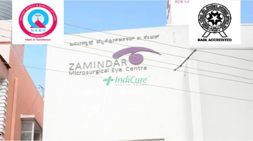
Zamindar's Microsurgical Eye Centre (ZMEC), which was founded in 1997, has been offering high-quality eye care to patients in and around Bangalore for the past 25 years. Their patients vouch for their services, and they are highly regarded for offering best-in-class eye treatment.
Allow the eye to recover. Don't do anything that could cause you to move your head. This includes activities such as cleaning or gardening, as well as moving swiftly and carrying anything heavy. You'll most likely need to take two to four weeks off work.
Sleeping on your side or even your front is preferable, but not on your back, as this will cause the bubble to shift away from the macular hole.
It's important to remember that the goal is to improve your eyesight in the damaged eye. A retinal detachment is a serious problem that can result in blindness. Your vision will very certainly never be as good as it was before your retinal detachment, even if surgery is successful.
"SF6" and "C3F8" are two of the most regularly used gasses. SF6 gas lasts about a month in the eye, while C3F8 gas lasts around two months. The most common gas utilized is SF6, with C3F8 gas reserved for more complicated retinal detachments and macular holes. For about a week, air remains in the eye.
There is no danger in watching TV or reading. For several weeks, your vision will be blurry or impaired. Following surgery, vision is frequently impaired. This will vary depending on the sort of procedure; for example, if a gas bubble is introduced into the eye, you may see the bubble's edge as it shrinks.
It's similar to a spirit level. Above this line, you will have vision, and below it, you will be blind. The line will gradually recede, with the viewing region growing larger and the black area shrinking until it is only a circle at the bottom of your vision, after which it will vanish.
Avoid jerky head movements and strenuous activities like lifting, cleaning, gardening, and so on.
Yes, you can definitely read after the surgery. Reading may be painful for a few days, but it is not harmful to the eyes.
With a single operation, the success rate for retinal detachment surgery is over 90%. This means that one out of every ten persons (10%) will require more than one operation. This is due to new tears forming in the retina or scar tissue forming in the eye, which contracts and pulls the retina off again.
Retinal surgery is normally painless and can be done while you are awake and relaxed. Outpatient eye surgery is now possible, thanks to technological advancements that have reduced the length of the procedure. You will be given anesthetic eye drops to numb your eyes before the treatment begins.
Sleeping on your side or even on your front is preferable, but not on your back, as this will cause the bubble to shift away from the macular hole.
While cost is an important factor, it is crucial to ensure that the chosen hospital and ENT surgeon have a good reputation, are board-certified, and have a proven track record in performing retinal detachment surgeries.
It is advisable to research thoroughly and read patient reviews before making a decision. Having said that, IndiCure does its own research to associate with surgeons who are highly experienced, skilled, and board-certified and are capable of delivering successful surgeries on international patients.
Traveling abroad for medical reasons may be challenging. With our experience of over a decade and working with the best surgeons and top hospitals in India, we help make your medical tour easier and safer for you. We will guide you at every step of the way and make end-to-end arrangements for your surgery, travel, and stay.
Ramandeep Dhaliwal
a month ago
I had great experience having rhinoplasty through Indicure. Dr. Ruchika from Indicure has helped me in finding best plastic surgeon, answering all my questions...
Read More
Joshua Archer
3 months ago
My name is Joshua Archer I'm from New Zealand, bay of plenty, kawerau I opted for the bypass surgery in January 2023 but planned it in advance for 28 September found IndiCure...
Read More
Kera Ren
8 months ago
Absolutely loved my experience with IndiCure - from first inquiring to meeting the surgeon pre op to my follow up post op. The surgeon was extremely approachable...
Read More
Andreana Paul
5 months ago
Had a wonderful experience. Visited India for my plastic surgery. From sending mails, airport pickup, comfortable accommodation and, to smooth hospital appointment booking...
Read More
Brandi Luce
5 months ago
I had the privilege of using Indicure's services for a cosmetic procedure that I had wanted for a long time but had always been apprehensive about. Ruchika helped me...
Read More
Jade M
3 years ago
Indicure Health Tours went above and beyond my expectations. They helped me with every aspect of my journey and were professional, kind and caring. I was...
Read More
The content on the website (www.indicure.com) is intended to be general information and is provided only as a service. All photographs on our website of before and after results are examples only, and do not constitute an implied or any other kind of certainty for the result of surgery.
Learn about IndiCure Health Tours' comprehensive editorial policy that strives to deliver trustworthy, helpful, relevant, accurate and people-first content on medical tourism in India.
It is not medical advice and should not be taken as medical advice. It should not be used to diagnose or treat a health condition and is in no way meant to be a substitute for professional medical care. You are advised to see a surgeon in person to assess what surgery may or may not accomplish for you.
It is also important to keep your expectations realistic and to understand that all surgical procedures carry risks and should never be taken lightly.